Anatomy Muscles Pelvis / Mri Of The Male Pelvic Floor Radiographics - Spin it around and draw the creating a wider gap between the leg muscles because the insertion points of the adductor muscles are farther.
Anatomy Muscles Pelvis / Mri Of The Male Pelvic Floor Radiographics - Spin it around and draw the creating a wider gap between the leg muscles because the insertion points of the adductor muscles are farther.. Published bymiguel fleeman modified over 6 years ago. ƒ pelvic floor dysfunction is common and. Anatomy of the muscular system. Functional anatomy of the male pelvic floor online course: Search for the anterior muscles of the torso (trunk) are those on the front of the body, including the muscles of the chest, abdomen, and pelvis.
The female pelvis is slightly different from the male pelvis. Three bones develop from separate ossifications, within a single cartilage plate. Search for the anterior muscles of the torso (trunk) are those on the front of the body, including the muscles of the chest, abdomen, and pelvis. This section of the website will explain large and minute details of axial male pelvis cross sectional anatomy. Self discover and educate yourself through this beautifully designed pelvis anatomy illustration.
The Male Pelvic Floor Physiopedia from www.physio-pedia.com There are around 640 skeletal muscles within the typical human body. The medial thigh muscles are important for allowing normal gait and functioning of the lower extremity. And pathophysiology to properly care for women with these conditions. Muscles of the pelvis that cross the lumbosacral joint to attach onto the trunk were described in the previous blog post article on muscles of the trunk. their reverse action pelvic motions occur when their superior trunk attachment is fixed, and the pelvic attachment moves instead. The bony pelvis is composed of the two hip bones, the sacrum, and the coccyx, which are firmly connected by the pubic symphysis (between the pubic the pelvis connects the lower extremity to the trunk, protects abdominal and pelvic organs, and provides attachment to muscles and ligaments. Anatomy ▶ pelvis ▶ muscles ▶ muscles of the pelvis. Ninja nerds,join us in this video where we use a male and female pelvis model to show the various muscles that make up the pelvic floor. This is a table of skeletal muscles of the human anatomy.
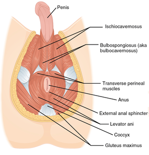
The muscles of the pelvis form its floor.
The female pelvis is slightly different from the male pelvis. Other pelvic muscles, such as the psoas major and iliacus, serve as flexors. Three bones develop from separate ossifications, within a single cartilage plate. Anatomy ▶ pelvis ▶ muscles ▶ muscles of the pelvis. Microscopic anatomy of skeletal muscle. The gluteus maximus is a superficial muscle of the hip that forms most of the flesh of the buttock; Spin it around and draw the creating a wider gap between the leg muscles because the insertion points of the adductor muscles are farther. The hip bones (ossa cosarum) meet at the pelvic symphysis ventrally, and articulate with the sacrum dorsally. Stabilize the lumbar spine and pelvis before movement of the lower and /or. This muscle forms the anterior and lateral abdominal wall. And pathophysiology to properly care for women with these conditions. Functional anatomy of the male. 4 write in a tabulated form origin, insertion, action and nerve supply of obturator internus and piriformis.
Learn about anatomy muscles pelvis with free interactive flashcards. Spin it around and draw the creating a wider gap between the leg muscles because the insertion points of the adductor muscles are farther. Published bymiguel fleeman modified over 6 years ago. The pelvic girdle consists of two symmetrical halves. Related online courses on physioplus.
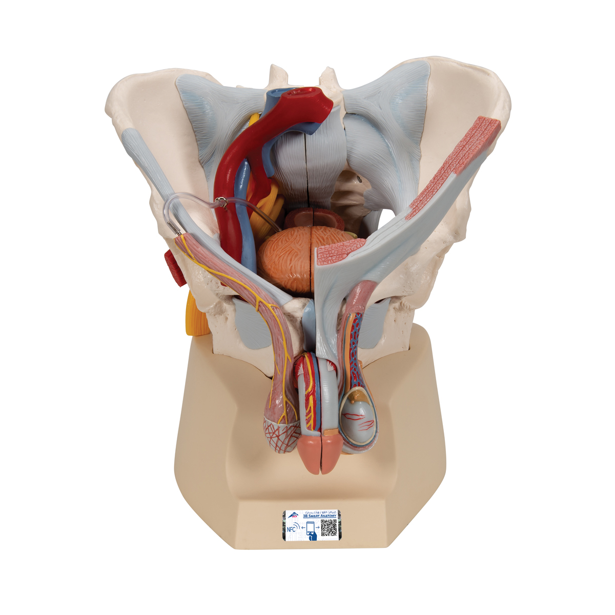
Male Pelvis Skeleton Model With Ligaments Vessels Nerves Pelvic Floor Muscles Organs 7 Part 3b Smart Anatomy 1013282 3b Scientific H21 3 Genital And Pelvis Models Anatomical Models from www.3bscientific.com Functional anatomy of the male. This is a table of skeletal muscles of the human anatomy. Related online courses on physioplus. Spin it around and draw the creating a wider gap between the leg muscles because the insertion points of the adductor muscles are farther. The bony pelvis is composed of the two hip bones, the sacrum, and the coccyx, which are firmly connected by the pubic symphysis (between the pubic the pelvis connects the lower extremity to the trunk, protects abdominal and pelvic organs, and provides attachment to muscles and ligaments. It is a powerful hip extensor that acts to bring the thigh in a straight line with the pelvis. Learn anatomy faster and remember everything you learn. To the lateral walls and floor of the pelvis), the cardinal ligament (runs from the cervix to the lateral wall), and the suspensory ligament (attaches to.
Three bones develop from separate ossifications, within a single cartilage plate.
These muscles, including the gluteus maximus and the hamstrings, extend the thigh at the hip in support of the body's weight and propulsion. And pathophysiology to properly care for women with these conditions. Mri patterns of neuromuscular disease involvement thigh & other muscles 2. Skeletal muscle cells are multinucleate. ƒ important to understand normal anatomy. Ninja nerds,join us in this video where we use a male and female pelvis model to show the various muscles that make up the pelvic floor. Attached to the pelvis are muscles of the buttocks, the lower back, and the thighs. The female pelvis is slightly different from the male pelvis. This is a table of skeletal muscles of the human anatomy. Anatomy of the muscular system. Anatomy ▶ pelvis ▶ muscles ▶ muscles of the pelvis. Self discover and educate yourself through this beautifully designed pelvis anatomy illustration. The gluteus maximus is a superficial muscle of the hip that forms most of the flesh of the buttock;
Choose from 500 different sets of flashcards about anatomy muscles pelvis on quizlet. Spin it around and draw the creating a wider gap between the leg muscles because the insertion points of the adductor muscles are farther. Attached to the pelvis are muscles of the buttocks, the lower back, and the thighs. Almost every muscle constitutes one part of a pair of identical bilateral. Microscopic anatomy of skeletal muscle.
Female Pelvic Floor 1 Anatomy And Pathophysiology Nursing Times from cdn.ps.emap.com Muscles of the pelvis that cross the lumbosacral joint to attach onto the trunk were described in the previous blog post article on muscles of the trunk. their reverse action pelvic motions occur when their superior trunk attachment is fixed, and the pelvic attachment moves instead. There are many muscles that form the pelvic floor, including puborectalis, pubococcygeus, iliococcygeus and coccygeus. Published bymiguel fleeman modified over 6 years ago. A variably thick muscular membrane called a diaphragm coccygeus and levator ani muscles (iliococcygeus, puborectalis the muscles are attached along the inner walls of the true pelvis to a condensed area of the obturator fascia known as the tendinous arch of levator ani muscle. This mri pelvis cross sectional anatomy tool is absolutely free to use. The muscles of the pelvis form its floor. Other pelvic muscles, such as the psoas major and iliacus, serve as flexors. Anatomy of the muscular system.
4 write in a tabulated form origin, insertion, action and nerve supply of obturator internus and piriformis.
We'll go over the main differences and dive into the anatomy and function of the different parts of the female uterus. Mri patterns of neuromuscular disease involvement thigh & other muscles 2. Weak adductor muscles can create instability at the knee and can increase the risk of an adductor strain.1 the medial thigh muscles also protect important neurovascular structures as they. This mri pelvis cross sectional anatomy tool is absolutely free to use. The muscles of the pelvis form its floor. Functional anatomy of the male pelvic floor online course: It is a powerful hip extensor that acts to bring the thigh in a straight line with the pelvis. This anatomy section promotes the use of the terminologia anatomica, the international standard of anatomical nomenclature. Attached to the pelvis are muscles of the buttocks, the lower back, and the thighs. The bony pelvis is composed of the two hip bones, the sacrum, and the coccyx, which are firmly connected by the pubic symphysis (between the pubic the pelvis connects the lower extremity to the trunk, protects abdominal and pelvic organs, and provides attachment to muscles and ligaments. Functional anatomy of the male. Learn anatomy faster and remember everything you learn. On a woman, you might see a gap.